High-Precision Anatomical Knee Joint (CT scan)
por IceGabrielito en MakerWorld
Boost MeWant more ANATOMY MODELS? Boost this model 🦴. 🩻Overview🩻. This high-precision 3D model is a faithful replica of the human knee joint, created through a rigorous anatomical and digital workflow. The skeletal structures were directly extracted and reconstructed from real CT scans to…
MakerWorld entrega los archivos a usuarios con cuenta gratuita
Licencia
Solo uso personalStandard Digital File License
Licencia estándar de archivos digitales: solo uso personal. No se permite compartir, revender ni subir el archivo a otros sitios, ni vender las impresiones.
Al descargar aceptas respetar la licencia y dar crédito a IceGabrielito.
- Categoría
- Educación
- Descargas
- 3,287
- Publicado
Sobre este modelo
Boost MeWant more ANATOMY MODELS? Boost this model 🦴
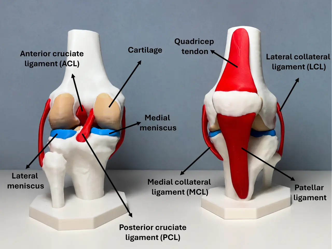
🩻Overview🩻 This high-precision 3D model is a faithful replica of the human knee joint, created through a rigorous anatomical and digital workflow. The skeletal structures were directly extracted and reconstructed from real CT scans to guarantee true-to-life bone morphology. The soft tissues—including ligaments, menisci, and articular cartilage—were then meticulously 3D modeled by hand, based on in-depth studies of MRI scans and anatomical literature. It serves as a premium medical-grade educational tool, perfect for students, orthopedic professionals, physical therapists, or 3D printing enthusiasts. Included Anatomical Structures The model features all critical bony and soft tissue structures necessary to understand the complex biomechanics of the knee:
- 🦴Skeletal Structure (CT Extracted): Femur, Tibia, Fibula, and Patella (kneecap), featuring accurate reproductions of condyles, epicondyles, and articular surfaces.
- 🔗Ligamentous Apparatus (MRI Modeled): - Cruciate Ligaments (Anterior Cruciate Ligament - ACL & Posterior Cruciate Ligament - PCL).
- Collateral Ligaments (Medial Collateral Ligament - MCL & Lateral Collateral Ligament - LCL).
- Patellar System (including the patellar tendon/ligaments).
- 🦵Cartilage & Cushioning (MRI Modeled): - Menisci (Medial and Lateral menisci).
- Articular Cartilage covering both the femoral and tibial contact surfaces.
Intended Use
- Education & Didactics: An excellent tactile and visual aid for anatomy classrooms, medical schools, and healthcare training.
- Patient Communication: Ideal for orthopedic clinics and physiotherapy practices to visually explain injuries (e.g., ACL tears, meniscus damage, osteoarthritis) and surgical procedures to patients.
- Advanced 3D Printing Showcase: A great project to showcase your printer's capability with complex, high-detail organic geometries.
💡 Print Settings & Recommendations
- Material: This model has been fully designed and optimized for PLA. No flexible filaments are required, making it easy and reliable to print on any standard 3D printer.
- Optimized for Single AMS (4 Colors): The model is structured to use exactly 4 colors, making it perfectly compatible with a single Bambu Lab AMS unit. To maximize visual contrast and educational value, the recommended color mapping is: - Bone White: Skeletal structures (Femur, Tibia, Fibula, Patella)
- Red: Ligaments
- Blue: Menisci
- Beige: Articular Cartilage
Disclaimer: This model is intended strictly for educational, illustrative, and hobbyist purposes. It is not a clinical tool and does not replace professional medical advice, diagnosis, or treatment.
Descripción original del autor, publicada en MakerWorld.